3D Imaging
High-resolution 3D Imaging for Intra-Operative & CT-Guided Procedures

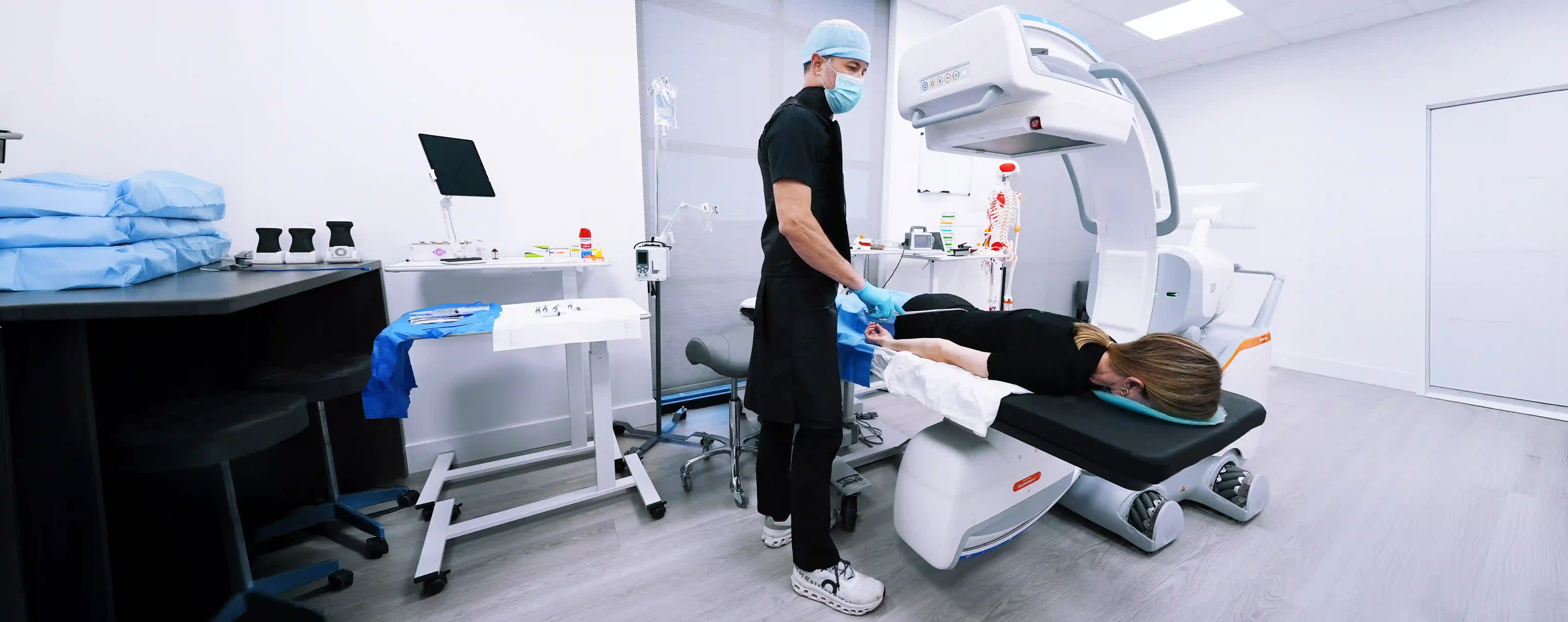
We have implemented the new Ciartic Move 3D imaging system, which is the most advanced imaging system of its kind that provides ultra-high-resolution 3D imaging with robotically guided movements, overcoming many of the limits of 2D image slices and using self-navigating capabilities to reduce the need for manual operation while also improving workflow during orthopedic procedures. The Siemens 3D Ciartic Move is a low-profile mobile imaging device that enables 3D navigation with robotic holonomic mechanisms to allow floating-like movements around a surgical field. This technology is the most advanced robotic 3D C-arm cone beam scanner that exists for interventional procedures to see targets and approaches in the highest resolution.
The Ciartic Move mobile 3D imaging system introduces a new era of mobile imaging and advances a wide array of intricate and complex minimally-invasive procedures and interventions in spine, orthopedics, and trauma by bringing together the most advanced technologies from 3D computed tomography (CT) with 3D navigation and needle guidance, metal artefact reduction and hardware detection, high-resolution fluoroscopy and magnification, digital subtraction angiography (DSA), low-dose radiation settings, and self-driving robotic guidance with automated positioning all at the touch of a button. As both a doctor and an engineer, Dr. McMurtrey is leading the way to combine the best of these fields to provide the most advanced and most minimally-invasive care possible.
We have already implemented the system in numerous procedures (see example videos below), and we are gaining new insights into the nuances of each injury and unique perspectives in minimally-invasive approaches to the critical features and pain triggering points of each injury, including spinal pars defects, sacroiliac dysfunction, spino-sacral anomalies, cranio-cervical instabilities, ligament destabilization, cartilage injuries and arthritic joint pathologies, degenerative disc defects and facet arthropathy affecting nerve roots and branches. In just the first week of implementing the system, it has enabled clear visualization of several complex problems that MRI imaging had been unable to fully clarify, including stress fractures in the spine and non-union fractures of the scaphoid bone in the wrist and of the sesamoid, metatarsal, and navicular bones in the foot, as well as complex bone spurs, synovial cysts, and subchondral bone cysts with arthritis that can be patched with orthobiologic scaffolding including patient’s own platelet-rich fibrin (PRF), bone marrow stem cells, and other regenerative agents. Furthermore, this technology is extremely valuable for confirming needle placement in small joints such as the atlanto-axial joints, atlanto-occipital joints, and facet joints of the spine.
This advanced 3D imaging further enables us to repair the underlying injuries as directly as possible with minimally-invasive image-guided procedures that let us see and target many types of orthopedic and neurologic problems from every angle. We are one of the only clinics that take a 3D tissue engineering approach to injuries, combining mesenchymal stem cells with biomaterial scaffolding architecture. When implemented with various types of spatially compartmentalized signaling factors, this will enable the greatest effects in tissue reconstruction, and this approach creates a potent combination of regenerative agents packed into a biocompatible patch that can integrate into the injured tissue defects and help stimulate repair to reconstruct proper tissue architecture. The final piece of the puzzle is then how to place these regenerative patches at injury sites in the least invasive way possible, and the Ciartic Move imaging system enables 3D image-guidance in minimally-invasive procedures to target injuries, lesions, cysts, defects, tears, fissures, and fractures with the highest resolution visualization. The Ciartic Move device is thus a powerful tool to help find and target sources of pain and directly treat a variety of injuries with minimally-invasive techniques that promote long-term recovery and more regenerative options to help repair tissue injuries and defects directly, thus leading us into the future of regenerative interventions.
-----
ASOI © 2025 All Rights Reserved